UCLA Core Technology Centers have created a new database of campus resources at http://www.cores.ucla.edu/
The site contains descriptions and contact information for each core, as well as a new searchable database of services and instrumentation (courtesy of the UCLA-DOE Institute Bioinformatics and Computational Core Technology Center).
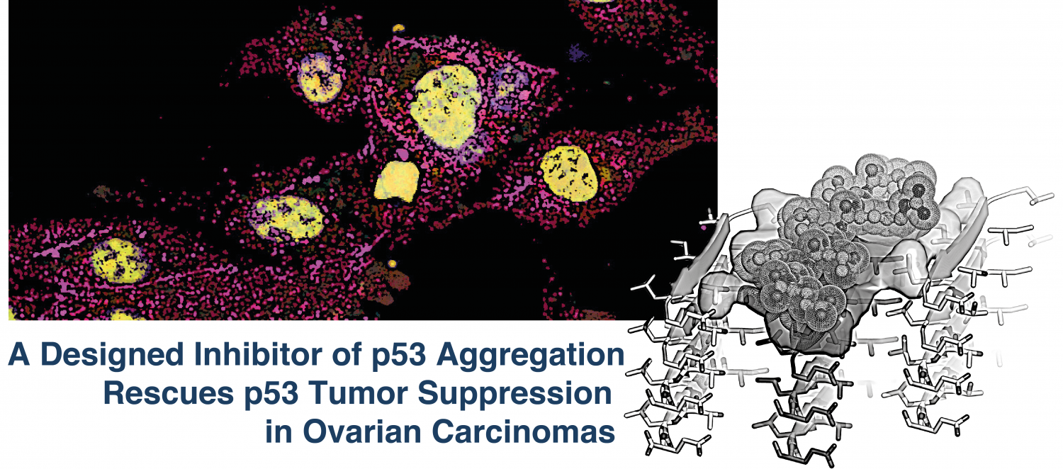
Approach borrows from technique of inhibiting protein fibers that cause Alzheimer’s and Parkinson’s diseases
* High-grade serous ovarian cancers are aggressive tumors, and most patients relapse despite standard therapy
* UCLA researchers developed a peptide that penetrates cancer cells and stops their growth by restoring the protective function of the p53 protein
* Scientists hope to test this therapeutic approach to treat women with ovarian cancer in the near future
UCLA scientists have developed a promising novel method to treat gynecologic tumors. The approach focuses on a protein called p53, which is commonly mutated in women who have high-grade serous ovarian cancer, the deadliest form of reproductive cancer. In many women with the disease, the cancer is very advanced by the time it is diagnosed and is therefore difficult to treat.
https://www.doe-mbi.ucla.edu/wp-content/uploads/2016/01/DOE_8-1.png27876317DOE-MBI Staffhttps://www.doe-mbi.ucla.edu/wp-content/uploads/2021/07/DOE3_newlogo4.jpgDOE-MBI Staff2016-01-06 23:19:242021-08-26 18:41:40UCLA Scientists Test New Strategy That Could Help Fight Ovarian Cancer
Published in the Journal of Biological Chemistry, the work of Dr. Lorena Saelices and coworkers under the supervision of Prof. David Eisenberg establishes a novel therapeutic strategy for transthyretin amyloidosis. The team identified two segments of transthyretin that are responsible for the protein aggregation and determined the structure of their fibrils. The fibril structures were used to design peptide inhibitors that block self-association of the two segments, and hinder transthyretin amyloid formation. The research published in December, 2015 opens a new approach to tackle the life-threatening transthyretin amyloidosis.
Abstract of the manuscript:
The tetrameric thyroxine-transport protein transthyretin (TTR) forms amyloid fibrils upon dissociation and monomer unfolding. The aggregation of transthyretin has been reported as the cause of the life-threatening transthyretin amyloidosis. The standard treatment of familial cases of TTR amyloidosis has been liver transplantation. Although aggregation-preventing strategies involving ligands are known, understanding the mechanism of TTR aggregation can lead to additional inhibition approaches. Several models of TTR amyloid fibrils have been proposed, but the segments that drive aggregation of the protein have remained unknown. Here we identify beta-strands F and H as necessary for TTR aggregation. Based on the crystal structures of these segments, we designed two non-natural peptide inhibitors that block aggregation. This work provides the first characterization of peptide inhibitors for TTR aggregation, establishing a novel therapeutic strategy
https://www.doe-mbi.ucla.edu/wp-content/uploads/2015/12/ttr_web2-1-copy-1.jpg10522400DOE-MBI Staffhttps://www.doe-mbi.ucla.edu/wp-content/uploads/2021/07/DOE3_newlogo4.jpgDOE-MBI Staff2015-12-01 22:47:572015-12-01 22:47:57Uncovering the Mechanism of Aggregation of Human Transthyretin
The Legacy Award honors outstanding MBI faculty mentors who have worked to inspire, encourage and guide the next generation of scientists and thought leaders.Professor Eisenberg is a Distinguished Professor of Chemistry and Biochemistry and of Biological Chemistry, an HHMI investigator, and the inaugural Chair Holder of the Paul D. Boyer Professorship in Molecular Biology Biochemistry. He served as director of the UCLA-DOE Institute for Genomics and Proteomics from 1993 to 2014.
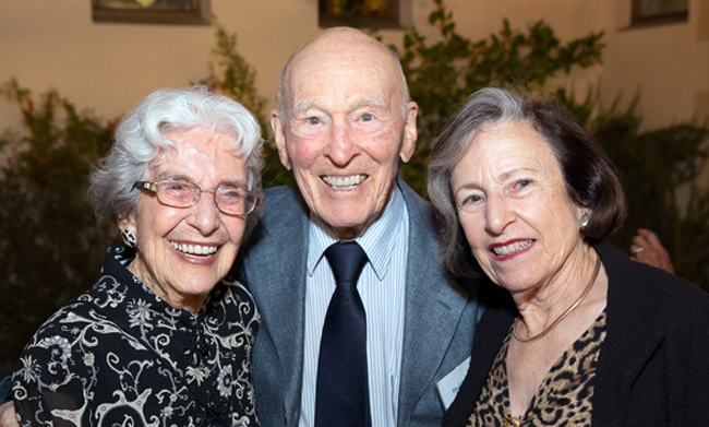
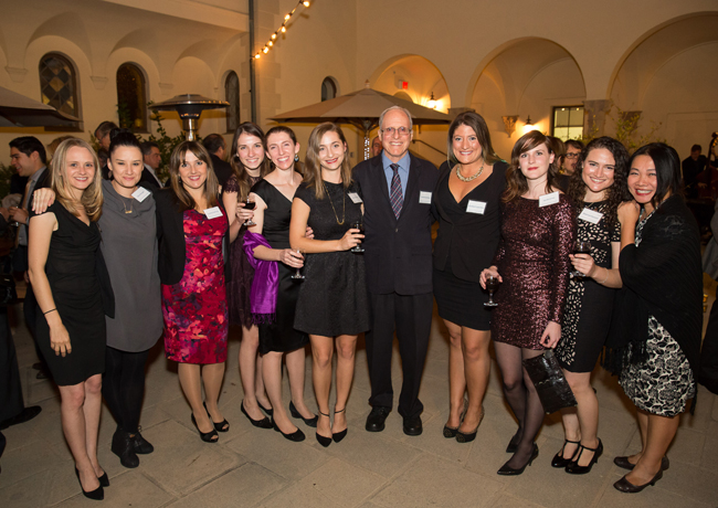
Nobel Laureate and founding MBI Director Paul D. Boyer and Lyda Boyer attended the event which was held at the UCLA Hershey Hall Grand Salon.
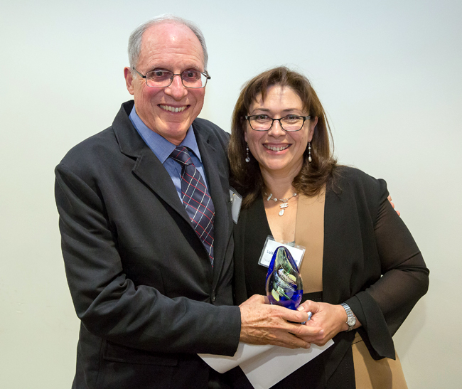
Professor David Eisenberg was presented the Legacy Award by MBI Director Professor Luisa Iruela-Arispe.
Left to right: Lyda Boyer, Professor Paul Boyer, and Lucy Eisenberg.
Members of Prof. Eisenberg’s group pose for a photo at the event (left to right): Dr. Nicole Wheatley, Dr. Alice Soragni, Dr. Lorena Saelices, Jeannette Bowler, Dr. Rebecca Nelson, Pascal Krotee, Prof. David Eisenberg, Elizabeth Guenther, Sarah Griner, Shannon Essewein, and assistant Cindy Chau.
Left to right: Professor Sabeeha Merchant (Chemistry & Biochemistry), Professor Edward De Robertis and Ana De Robertis (Biological Chemistry).
https://www.doe-mbi.ucla.edu/wp-content/uploads/2015/11/MBI-Awards-Dinner-small-1.jpg11082DOE-MBI Staffhttps://www.doe-mbi.ucla.edu/wp-content/uploads/2021/07/DOE3_newlogo4.jpgDOE-MBI Staff2015-11-18 23:32:342021-08-26 19:05:13Professor David Eisenberg was awarded the Legacy Award at the 2nd Annual UCLA Molecular Biology Institute (MBI) Awards Dinner on November 12th.
A collaboration between Bowie Laboratory and a research team led by Tae-Young Yoon of the Department of Physics at the Korea Advanced Institute of Science and Technology has employed the use of a magnetic tweezer to “quantitatively map the folding energy landscape, the folding kinetic rate, and folding intermediates of a membrane protein in a membrane environment for the first time.” (Phys.org)
Members of Bowie lab have provided the following abstract and external links:
Membrane proteins are designed to fold and function in a lipid membrane, yet folding experiments within a native membrane environment are challenging to design. Here we show that single molecule forced unfolding experiments can be adapted to study helical membrane protein folding under native-like bicelle conditions. Applying force using magnetic tweezers, we find that a transmembrane helix protein, E. coli rhomboid protease GlpG, unfolds in a highly cooperative manner, largely unraveling as one physical unit in response to mechanical tension above 25 pN. Considerable hysteresis is observed, with refolding occurring only at forces below 5 pN. Characterizing the energy landscape reveals only modest thermodynamic stability (DG = 6.5 kBT) but a large unfolding barrier (21.3 kBT) that can maintain the protein in a folded state for long periods of time (t1/2 ~3.5 hrs). The observed energy landscape may have evolved to limit the existence of troublesome partially unfolded states and impart rigidity to the structure.
https://www.doe-mbi.ucla.edu/wp-content/uploads/2015/10/Untitled-1-1.png482371Michael Collazohttps://www.doe-mbi.ucla.edu/wp-content/uploads/2021/07/DOE3_newlogo4.jpgMichael Collazo2015-10-19 01:00:322015-10-19 01:00:32Mapping the energy landscape for second stage folding of a single membrane protein
“Protein aggregates associated with neurodegenerative disease have stubbornly resisted researchers’ efforts to get a good look at them. They refuse to crystallize well or yield to standard spectroscopic techniques. Now, advances in electron microscopy methods are forcing these molecules to give up their secrets. In the September 9 Nature, researchers led by David Eisenberg at the University of California, Los Angeles, and Tamir Gonen at the Howard Hughes Medical Institute’s Janelia Research Campus in Ashburn, Virginia, offer the closest look yet at the core of α-synuclein aggregates. The researchers made microscopic crystals from the peptides and used a relatively new technique called micro-electron diffraction to map them down to atomic resolution. Commenters called the work a tour de force.
‘[These] structures are the first to be determined by micro-electron diffraction from a molecule of previously unknown structure,’ noted Yifan Cheng at the University of California, San Francisco, in an accompanying Nature commentary. Others were similarly impressed. ‘To obtain a structure of this quality from a peptide material with such tiny crystals is a remarkable feat, and will probably serve as the model for many other studies,’ Gregory Petsko at Weill Cornell Medical College in New York told Alzforum. Tim Bartels at Brigham and Women’s Hospital, Boston, agreed, ‘The resolution here is unprecedented.’”
Important three-dimensional spatial features such as depth and surface concavity can be difficult to convey clearly in the context of two-dimensional images. In the area of macromolecular visualization, the computer graphics technique of ray-tracing can be helpful, but further techniques for emphasizing surface concavity can give clearer perceptions of depth. The notion of diffusion accessibility is well-suited for emphasizing such features of macromolecular surfaces, but a method for calculating diffusion accessibility has not been made widely available. Here we make available a web-based platform that performs the necessary calculation by solving the Laplace equation for steady state diffusion, and produces scripts for visualization that emphasize surface depth by coloring according to diffusion accessibility. The URL is http://services.mbi.ucla.edu/DiffAcc/.
https://www.doe-mbi.ucla.edu/wp-content/uploads/2015/08/mhc_after_sml-1.jpg180180DOE-MBI Staffhttps://www.doe-mbi.ucla.edu/wp-content/uploads/2021/07/DOE3_newlogo4.jpgDOE-MBI Staff2015-10-01 00:40:092015-10-01 00:40:09Diffusion accessibility as a method for visualizing macromolecular surface geometry.
In a new PNAS article, members of the UCLA-DOE Institute, Andrew AhYoung, Pascal Egea, and Joseph Loo, together with Jiansen Jiang, Jiang Zhang, and Hong Zhou, have offered further elucidation on the structure and function of the conserved SMP domains in the ERMES complex.
The new article asserts the roles played by conserved SMP domains of ERMES in the tethering of mitochondrial and endoplasmic reticulum membranes and the essential exchange of phosholipids, such as phosphatidylcholines, between the two organelles.