IGP Core
Protein Expression Technology
CORE MANAGER: MARK ARBING
Phone:
(310) 206-2871
Email:
marbing@mbi.ucla.edu
Boyer Hall 103, 611 Young Drive East, Los Angeles, CA 90095

IGP Core
Phone:
(310) 206-2871
Email:
marbing@mbi.ucla.edu
Boyer Hall 103, 611 Young Drive East, Los Angeles, CA 90095
The center was founded in 1994 to facilitate the expression and purification of proteins for structure/function studies. The core provides support in all aspects of protein expression from cloning through expression optimization and protein purification. The center is a UCLA-DOE Institute for Genomics and Proteomics facility but is open to all researchers.
The evaluation of many expression constructs can be a crucial step in obtaining a protein sample suitable for further characterization. Recent advances in cloning and protein expression based on technologies developed by structural genomics consortia have resulted in the ability to evaluate tens or hundreds of expression constructs rapidly and economically. We have adapted these techniques to allow for a medium throughput approach suitable for the hypothesis-driven projects of academic research groups. Our resources allow rapid analysis of conditions that yield well-behaved soluble protein for downstream biochemical and biophysical studies.
RATES
Rates are for experiments performed using our standardized protocols. Customized projects are welcome; contact us for questions about experimental design and pricing.
|
Service |
UC-rate |
External rate |
|
Protein Expression/Purification |
||
|
Bacterial transformation (into available expression strains) |
$74.64 |
$149.36 |
|
Large-scale cell growth (1 L) |
$74.64 |
$149.36 |
|
12 L Fermentation (with BioEngineering NLF22 Bioreactor) |
$645.45 |
$1,291.55 |
|
96-well protein expression and purification; includes transformation (maximum number of strain plasmid combinations is 8) of expression plasmids into available expression strains, IPTG-induction of protein expression, cell harvesting and lysis with buffers containing solubility promoting additives, Ni-NTA affinity purification, SDS-PAGE analysis. |
$1,463.03 |
$2,927.52 |
|
Large-scale (bacterial); 1 L cell growth with user-supplied transformed expression strain, Ni-NTA affinity purification, SDS-PAGE analysis, protein concentration and buffer exchange by dialysis. |
$1,347.53 |
$2,696.40 |
|
Additional chromatography steps |
$156.75 |
$313.66 |
|
Protein concentration/Dialysis |
$39.28 |
$78.60 |
|
Data analysis/troubleshooting/protein construct design (per hour plus reagent cost) |
$82.80 |
$165.68 |
*User supplies the template or we clone from our genomic DNA collection for an additional cost.
CLONING AND CONSTRUCT DESIGN
The PETC clones genes directly from an organism, library, or synthetic DNA using any common cloning techniques although our preferred method is based on Gibson assembly. We maintain a supply of approximately 65 bacterial genomes to use as PCR templates; however most current projects use third party gene synthesis vendors to supply gene fragments or sequence-verified expression clones. A variety of bioinformatics tools (PSIPRED, DISOPRED, Phyre, AlphaFold, SignalP, etc) are used to guide the design of expression constructs.
EXPRESSION SYSTEMS
Bacterial expression systems used by the PETC include a variety of inducible protein expression vectors (pET and pBAD), fusion systems (SUMO/His6/MBP/Thioredoxin) for increased solubility and affinity chromatography, and vectors to co-express chaperones or rare tRNAs to promote folding and increase recombinant protein yields. Eukaryotic expression systems, yeast (S. cerevisiae and P. pastoris) or insect cells (Sf9) use constitutive or inducible promoters and allow for secreted or cytoplasmic expression of target proteins.
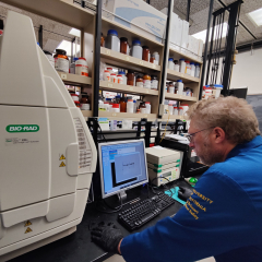
PROTEIN EXPRESSION SCREENING
Protein expression screening uses bacterial (E. coli) and yeast (P. pastoris) expression hosts. A ShelLab SI6R-HS shaking incubator allows growth of up to 1152 cultures in 96-well format to high culture densities. The high cell densities allow microgram amounts of recombinant protein to be produced in the small culture volumes used in 96-well plates. Cell lysis is performed in 96-well format and SDS-PAGE analysis is used to evaluate target protein expression. Batch purification of protein from the soluble fraction is used to assess the ability of the target protein to bind affinity purification resins. Cell growth in 96-well format allows parallel investigation of parameters critical for obtaining high yields of recombinant target proteins. Parameters explored include: expression strains, media, growth and/or induction temperature, coexpression with accessory plasmids (pLysS, pRIL, pRARE, etc), and lysis buffers.
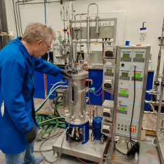
LARGE SCALE FERMENTATION AND CELL LYSIS
After suitable protein expression/solubility conditions have been determined this information is used to scale up cell growth to increase recombinant protein yields. The PETC is equipped with New Brunswick Innova refrigerated shaking incubators and three BioEngineering NLF22 fermentors (working volumes of ~12L.) The PETC is equipped with a sonicator, French press, and two Avestin Emulsiflex C-3 high pressure cell homogenizers. The Emulsiflex instruments are particularly well suited for efficient high-throughput cell lysis with a throughput of up to 3L of lysate per hour.
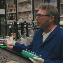
PROTEIN PURIFICATION
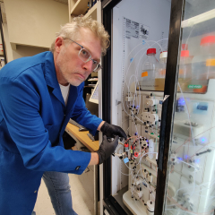
Using predetermined information on protein expression and solubility from small-scale experiments, recombinant proteins are purified in large-scale using Bio-Rad NGC chromatography systems. Chromatography methods include: affinity (GST, amylose, metal chelate, heparin, dye, etc.), ion exchange (anion or cation), hydrophobic interaction (HIC), and size exclusion chromatography. If required fusion proteins and/or affinity purification tags can be proteolytically cleaved and removed at this stage.
One of the Bio-Rad NGC systems is equipped with air sensors, sample pump, and multiple column valves allowing completely automated purification of up to seven samples by multiple chromatographic methods. The instrument is configured to allow unattended multi-step purification of samples by different chromatographic methods, e.g. affinity chromatography followed by a desalting or size exclusion chromatography step. The second NGC system has a four wavelength UV detector allowing four distinct wavelengths to be monitored at the same time allowing simultaneous detection of protein, nucleic acids, and chromophores.
FLUORESCENCE ACTIVATED CELL SORTING (FACS) AND YEAST DISPLAY
The Bio-Rad S3 Cell Sorter is an easy to use automated cell sorter accessible to both novices and experts. The system is equipped with two lasers (488 and 561 nm) and four fluorescence detectors plus forward- and side-scatter detectors.
The PETC has constructed a yeast DARPin display library for identification of high-affinity protein binders and uses the S3 instrument for enrichment of high-affinity binders from naïve libraries. These binders can be used for structural biology applications such as crystallization chaperones and as adaptors for cryoEM imaging scaffolds; they have additional uses as tools for a variety of biochemical and cell-based assays. The PETC has produced low nanomolar affinity binders for academic labs and the biotech industry.
CRYSTALLOGRAPHY
Structural studies of target proteins can be coordinated with the X-Ray and EM Structure Determination Core.
INSTRUMENTATION
USER AGREEMENT and SOPs
Our services have greatly benefitted the local research community leading to numerous publications. The most recent are listed below with names of PETC staff members in bold.
Vadivel K, Zaiss AK, Kumar Y, Fabian FM, Ismail AEA, Arbing MA, Buchholz WG, Velander WH, Bajaj SP. 2020. Enhanced Antifibrinolytic Efficacy of a Plasmin-Specific Kunitz-Inhibitor (60-Residue Y11T/L17R with C-Terminal IEK) of Human Tissue Factor Pathway Inhibitor Type-2 Domain1. J Clin Med. 9:3684.
Valliere MA, Korman TP, Arbing MA, Bowie JU. 2020. A bio-inspired cell-free system for cannabinoid production from inexpensive inputs. Nat Chem Biol. 16:1427-1433.
Satagopan S, North JA, Arbing MA, Varaljay VA, Haines SN, Wildenthal JA, Byerly KM, Shin A, Tabita FR. 2019. Structural Perturbations of Rhodopseudomonas palustris Form II RuBisCO Mutant Enzymes That Affect CO2 Fixation. Biochemistry. 58:3880-3892.
Tuukkanen AT, Freire D, Chan S, Arbing MA, Reed RW, Evans TJ, Zenkeviciutė G, Kim J, Kahng S, Sawaya MR, Chaton CT, Wilmanns M, Eisenberg D, Parret AHA, Korotkov KV. 2018. Structural Variability of EspG Chaperones from Mycobacterial ESX-1, ESX-3, and ESX-5 Type VII Secretion Systems. J Mol Biol. 431:289-307.
Min D, Jefferson RE, Qi Y, Wang JY, Arbing MA, Im W, Bowie JU. 2018. Unfolding of a ClC chloride transporter retains memory of its evolutionary history. Nat Chem Biol. 14:489-496.
Bajaj RA, Arbing MA, Shin A, Cascio D, Miallau L. 2016. Crystal structure of the toxin Msmeg_6760, the structural homolog of Mycobacterium tuberculosis Rv2035, a novel type II toxin involved in the hypoxic response. Acta Crystallogr F Struct Biol Commun. 72:863-869.
Min D, Arbing MA, Jefferson RE, Bowie JU. 2016. A simple DNA handle attachment method for single molecule mechanical manipulation experiments. Protein Sci. 25:1535-44.
Wagner JM, Chan S, Evans TJ, Kahng S, Kim J, Arbing MA, Eisenberg D, Korotkov KV. 2016. Structures of EccB1 and EccD1 from the core complex of the mycobacterial ESX-1 type VII secretion system. BMC Struct Biol. 16:5.
Taylor ND, Garruss AS, Moretti R, Chan S, Arbing MA, Cascio D, Rogers JK, Isaacs FJ, Kosuri S, Baker D, Fields S, Church GM, Raman S. 2016. Engineering an allosteric transcription factor to respond to new ligands. Nat Methods. 13:177-83.
Varaljay VA, Satagopan S, North JA, Witte B, Dourado MN, Anantharaman K, Arbing MA, Hoeft McCann S, Oremland RS, Banfield JF, Wrighton KC, Tabita FR. 2015. Functional metagenomic selection of RubisCO from uncultivated bacteria. Environ Microbiol. 18:1187-99.
Rodriguez JA, Ivanova MI, Sawaya MR, Cascio D, Reyes FE, Shi D, Sangwan S, Guenther EL, Johnson LM, Zhang M, Jiang L, Arbing MA, Nannenga BL, Hattne J, Whitelegge J, Brewster AS, Messerschmidt M, Boutet S, Sauter NK, Gonen T, Eisenberg DS. Structure of the toxic core of α-synuclein from invisible crystals. 2015. Nature. 525: 486-90.
Leibly DJ, Arbing MA, Pashkov I, DeVore N, Waldo GS, Terwilliger TC, Yeates TO. A Suite of Engineered GFP Molecules for Oligomeric Scaffolding. 2015. Structure. 23: 1754-68.
Bobik TA, Morales EJ, Shin A, Cascio D, Sawaya MR, Arbing M, Yeates TO, Rasche, M. 2014. Structure of the methanofuran/methanopterin biosynthetic enzyme MJ1099 from Methanocaldococcus jannaschii. Acta Cryst. F70: 1472-1479.
Satagopan S, Chan S, Perry LJ, Tabita FR. 2014. Structure-function studies with the unique hexameric form II ribulose-1,5-bisphosphate carboxylase/oxygenase (Rubisco) from Rhodopseudomonas palustris. J Biol Chem. 289: 21433-50.
Arbing MA, Chan S, Harris L, Kuo E, Zhou TT, Ahn CJ, Nguyen L, He Q, Lu J,Menchavez PT, Shin A, Holton T, Sawaya MR, Cascio D, Eisenberg D. 2013. Heterologous xxpression of mycobacterial Esx complexes in Escherichia coli for structural studies is facilitated by the use of maltose binding protein fusions. PLoS One. 8: e81753.
Miallau L, Jain P, Arbing MA, Cascio D, Phan T, Ahn CJ, Chan S, Chernishof I, Maxson M, Chiang J, Jacobs WR Jr, Eisenberg DS. 2013. Comparative proteomics identifies the cell-associated lethality of M. tuberculosis RelBE-like toxin-antitoxin complexes. Structure. 21: 627-37.
Arbing MA, Chan S, Shin A, Phan T, Ahn CJ, Rohlin L, Gunsalus RP. 2012. Structure of the surface layer of the methanogenic archaean Methanosarcina acetivorans. Proc Natl Acad Sci USA. 109: 11812-7.
Urzica EI, Adler LN, Page MD, Linster CL, Arbing MA, Casero D, Pellegrini M, Merchant SS, Clarke SG. 2012. Impact of oxidative stress on ascorbate biosynthesis in Chlamydomonas via regulation of the VTC2 gene encoding a GDP-L-galactose phosphorylase. J Biol Chem. 287:14234-45.
Arbing MA, Kaufmann M, Phan T, Chan S, Cascio D, Eisenberg D. 2010. The crystal structure of the Mycobacterium tuberculosis Rv3019c-Rv3020c ESX complex reveals a domain-swapped heterotetramer. Protein Sci. 19: 1692-703.
Chan S, Giuroiu I, Chernishof I, Sawaya MR, Chiang J, Gunsalus RP, Arbing MA, Perry LJ. 2010. Apo and ligand-bound structures of ModA from the archaeon Methanosarcina acetivorans. Acta Cryst. F66: 242-50.
Miallau L, Faller M, Chiang J, Arbing M, Guo F, Cascio D, Eisenberg D. 2009. Structure and proposed activity of a member of the VapBC family of toxin-antitoxin systems. VapBC-5 from Mycobacterium tuberculosis. J Biol Chem. 284: 276-83.
Tabita FR, Hanson TE, Li H, Satagopan S, Singh J, Chan S. 2007. Function, structure, and evolution of the RubisCO-like proteins and their RubisCO homologs. Microbiol Mol Biol Rev. 71: 576-99.
Choi B, Zocchi G, Canale S, Wu Y, Chan S, Perry LJ. 2005. Artificial allosteric control of maltose binding protein. Phys Rev Lett. 94: 038103.
Sherkhanov S, Korman TP, Chan S, Faham S, Liu H, Sawaya MR, et al. 2020. Isobutanol production freed from biological limits using synthetic biochemistry. Nat Commun. 11: 4292.
Liclican EL, Filler SG, Kaye J, Denny CT. 2019. Describing the dynamic translational science landscape through Core Voucher utilization. J Clin Transl Sci. 3: 105–112.
Sarafian TA, Yacoub A, Kunz A, Aranki B, Serobyan G, Cohn W, et al. 2019. Enhanced mitochondrial inhibition by 3,4-dihydroxyphenyl-acetaldehyde (DOPAL)-oligomerized α-synuclein. J Neurosci Res. 97: 1689–1705.
Chang ZL, Lorenzini MH, Chen X, Tran U, Bangayan NJ, Chen YY. 2018. Rewiring T-cell responses to soluble factors with chimeric antigen receptors. Nat Chem Biol. 14: 317–324.
Messina MS, Stauber JM, Waddington MA, Rheingold AL, Maynard HD, Spokoyny AM. 2018. Organometallic Gold(III) Reagents for Cysteine Arylation. J Am Chem Soc. 140: 7065–7069.
Campagna J, Vadivel K, Jagodzinska B, Jun M, Bilousova T, Spilman P, et al. 2018. Evaluation of an Allosteric BACE Inhibitor Peptide to Identify Mimetics that Can Interact with the Loop F Region of the Enzyme and Prevent APP Cleavage. J Mol Biol. 430: 1566–1576.
Zimmer RK, Ferrier GA, Kim SJ, Loo RRO, Zimmer CA, Loo JA. 2017. Keystone predation and molecules of keystone significance. Ecology. 98: 1710–1721.
He C, Hu X, Jung RS, Larsson M, Tu Y, Duarte-Vogel S, et al. 2017. Lipoprotein lipase reaches the capillary lumen in chickens despite an apparent absence of GPIHBP1. JCI Insight. 2: e96783.
Sarafian TA, Littlejohn K, Yuan S, Fernandez C, Cilluffo M, Koo BK, Whitelegge JP, Watson JB. Stimulation of synaptoneurosome glutamate release by monomeric and fibrillated α-synuclein. J Neurosci Res. 2017 Jan 24. doi: 10.1002/jnr.24024.
Decker CG, Wang Y, Paluck SJ, Shen L, Loo JA, Levine AJ, Miller LS, Maynard HD. 2016. Fibroblast growth factor 2 dimer with superagonist in vitro activity improves granulation tissue formation during wound healing. Biomaterials. 81:157-68.
Fristedt R, Martins NF, Strenkert D, Clarke CA, Suchoszek M, Thiele W, Schöttler MA, Merchant SS. The thylakoid membrane protein CGL160 supports CF1CF0 ATP synthase accumulation in Arabidopsis thaliana. 2015. PLoS One. 10: e0121658.
Fristedt R, Herdean A, Blaby-Haas CE, Mamedov F, Merchant SS, Last RL, Lundin B. PHOTOSYSTEM II PROTEIN33, a protein conserved in the plastid lineage, is associated with the chloroplast thylakoid membrane and provides stability to photosystem II supercomplexes in Arabidopsis. 2015. Plant Physiol. 167: 481-92.
Fristedt R, Williams-Carrier R, Merchant SS, Barkan A. A thylakoid membrane protein harboring a DnaJ-type zinc finger domain is required for photosystem I accumulation in plants. 2014. J Biol Chem. 289: 30657-67.
Fristedt R, Scharff LB, Clarke CA, Wang Q, Lin C, Merchant SS, Bock R. RBF1, a plant homolog of the bacterial ribosome-binding factor RbfA, acts in processing of the chloroplast 16S ribosomal RNA. 2014. Plant Physiol. 164: 201-15.
Hsieh SI, Castruita M, Malasarn D, Urzica E, Erde J, Page MD, Yamasaki H, Casero D, Pellegrini M, Merchant SS, Loo JA. 2013. The proteome of copper, iron, zinc, and manganese micronutrient deficiency in Chlamydomonas reinhardtii. Mol Cell Proteomics. 12: 65-86.
Sergeev ME, Morgia F, Javed MR, Doi M, Keng PY. 2013. Enzymatic radiofluorination: Fluorinase accepts methylaza-analog of SAM as substrate for FDA synthesis. J Mol Catal B Enzym. 97: 74–79.
Min AB, Miallau L, Sawaya MR, Habel J, Cascio D, Eisenberg D. 2012. The crystal structure of the Rv0301-Rv0300 VapBC-3 toxin—antitoxin complex from M. tuberculosis reveals a Mg2+ ion in the active site and a putative RNA-binding site. Protein Sci. 21: 1754–1767.
Teng PK, Anderson NJ, Goldschmidt L, Sawaya MR, Sambashivan S, Eisenberg D. 2012. Ribonuclease A suggests how proteins self-chaperone against amyloid fiber formation. Protein Sci. 21: 26–37.
Besserer GM, Nicoll DA, Abramson J, Philipson KD. 2012. Characterization and purification of a Na+/Ca2+ exchanger from an archaebacterium. J Biol Chem. 287: 8652-9.
Wang A, Zocchi G. 2011. Artificial modulation of the gating behavior of a K+ channel in a KvAP-DNA chimera. PLoS One. 6: e18598.
Chim N, Habel JE, Johnston JM, Krieger I, Miallau L, Sankaranarayanan R, Morse RP, Bruning J, Swanson S, Kim H, Kim CY, Li H, Bulloch EM, Payne RJ, Manos-Turvey A, Hung LW, Baker EN, Lott JS, James MN, Terwilliger TC, Eisenberg DS, Sacchettini JC, Goulding CW. 2011. The TB Structural Genomics Consortium: a decade of progress. Tuberculosis (Edinb). 91: 155-72.
Laganowsky A, Benesch JLP, Landau M, Ding L, Sawaya MR, Cascio D, Huang Q, Robinson CV, Horwitz J, Eisenberg D. 2010. Crystal structures of truncated alphaA and alphaB crystallins reveal structural mechanisms of polydispersity important for eye lens function. Protein Sci. 19: 1031–1043.
Li PP, Itoh N, Watanabe M, Shi Y, Liu P, Yang HJ, Kasamatsu H. 2009. Association of simian virus 40 vp1 with 70-kilodalton heat shock proteins and viral tumor antigens. J Virol. 83: 37-46.