
Protein crystal structure obtained at 2.9 Å resolution from injecting bacterial cells into an X-ray free-electron laser beam.
The Eisenberg lab led an international team of 22 scientists in obtaining a 2.9 angstrom resolution protein crystal structure by injecting bacterial cells into an X-ray free-electron laser beam. Their accomplishment was remarkable because unlike the ∼100,000 biological structures determined by X-ray crystallography to date, the macromolecule under study was not extracted from the cells that produced it.
The whole cell was injected in to the beam, with no attempt to isolate the crystals from the other cell components. Furthermore, the average size of the crystals was exceedingly small, approximately 1.5 x 1.0 x 0.5 microns, so it was impossible to collect single-crystal diffraction patterns using current state-of-the-art microfocus technology. The team found in this case that scattering from non-crystalline cell components such as the cell membrane and spores did not obscure diffraction from the crystals. The study suggests that as technology advances, it will become feasible to obtain high resolution images of smaller, less ordered systems as they exist in the cell –glimpsing life on the molecular level.
Sawaya MR, Cascio D, Gingery M, Rodriguez J, Goldschmidt L, Colletier JP, Messerschmidt MM, Boutet S, Koglin JE, Williams GJ, Brewster AS, Nass K, Hattne J, Botha S, Doak RB, Shoeman RL, DePonte DP, Park HW, Federici BA, Sauter NK, Schlichting I, Eisenberg DS. Protein crystal structure obtained at 2.9 Å resolution from injecting bacterial cells into an X-ray free-electron laser beam. Proc Natl Acad Sci U S A. 2014 Sep 2;111(35):12769-74. doi: 10.1073/pnas.1413456111. Epub 2014 Aug 18. PubMed PMID: 25136092; PubMed CentralPMCID: PMC4156696..
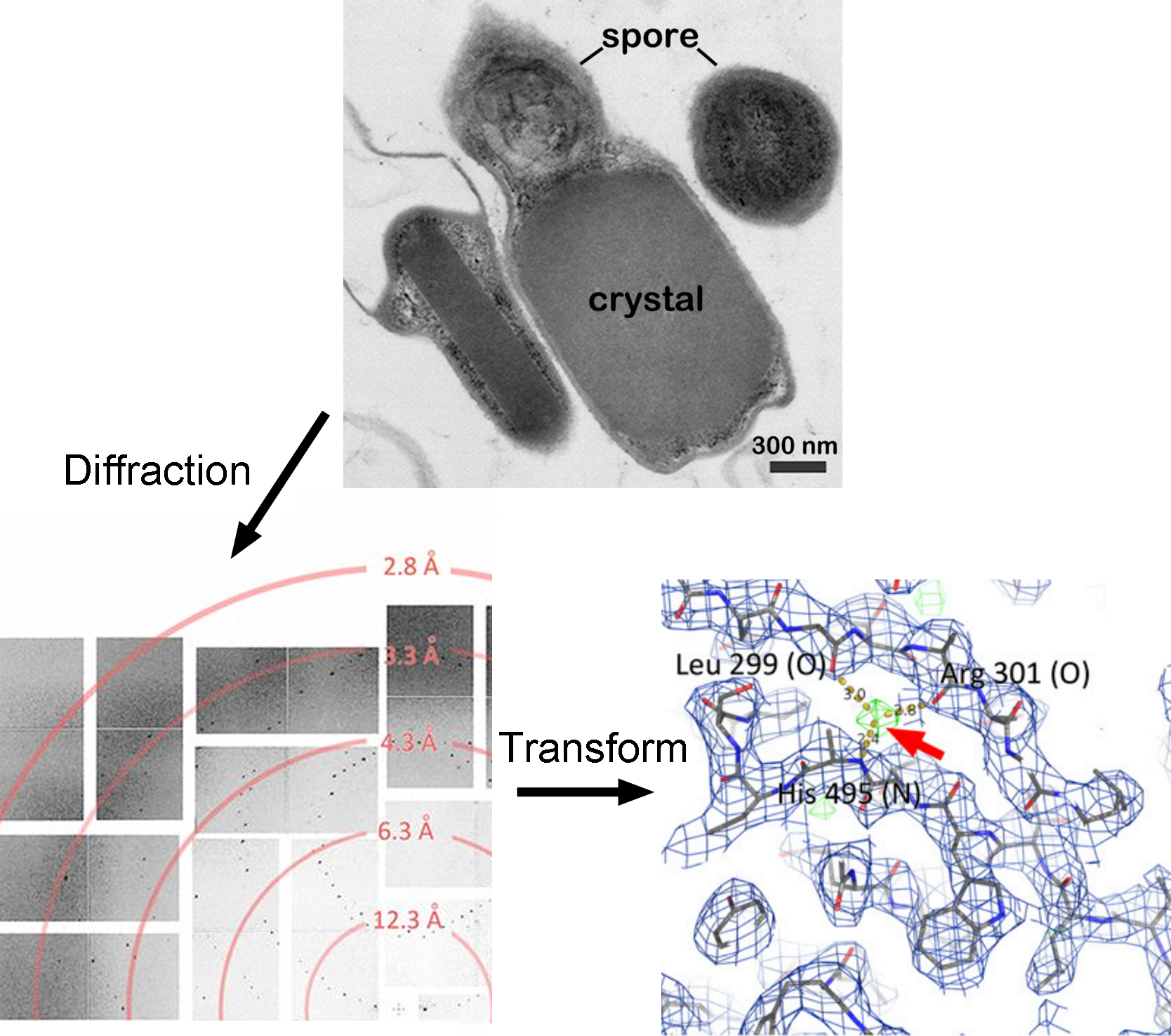
Top panel is an electron micrograph of Bacillus thuringiensis used in this study. The crystal occupies most of the cell volume. Diffraction from the cells (lowerleft) extends to 2.9 Å resolution. Lower right shows structural detail.