
Next APS Trip
April 17th to the 20th (2015)
International collaboration co-led by Prof. David Eisenberg elucidates the mechanism of safe storage and action of the potent human toxin MBP-1
Eosinophils are white blood cells that are part of the body’s innate immune defense against pathogens. Once an infection occurs, eosinophils are activated and migrate to the infected tissue where they contribute to kill the invading microorganisms (bacteria, viruses or helminths) by secreting a number of toxic proteins, including the Major Basic Protein (MBP-1).
While eosinophils are typically maintained at very low numbers in blood, in certain diseases, such as allergies, bronchial asthma, eosinophilic esophagitis or other eosinophilic syndromes, a highly increased number of cells is present. In these diseases, eosinophils can be aberrantly activated and infiltrate organs where, by releasing MBP-1 and other toxins, they can generate substantial tissue damage.
The Eisenberg lab co-led an international collaboration of scientists from over 10 different institutions to elucidate the details of how the powerful MBP-1 toxin is safely stored inside the eosinophil cell as well as the mechanism of its toxicity upon extracellular release. The findings were published this month in Molecular Cell.
Crowd Phasing
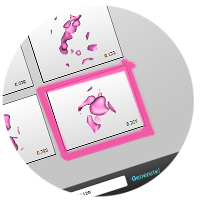 We have a new CrowdPhasing game that we would like everyone to try to play. It was begun briefly as part of a conference demonstration last month, and it was a promising start, but now we need to play the game in earnest to see how far it can progress. This one is distinctly different from previous games. This one is based on a *racemic* crystal, which simplifies the phase problem by allowing only two possible choices for each reflection (as you probably know). So far the experience is that this racemic game is a bit easier to make work than previous ones.
As usual, the purpose of the game is to try to drive the phases and their corresponding electron density maps to the correct solution, which should look like a very low resolution boundary of the unknown molecule. In the present case, we take as known information only that the unit cell should contain two copies of a small/compact cyclic polypeptide, whose molecular envelope should be relatively simple in shape at low resolution. Please join and play the game in the usual way. The name of this game is “Racemic peptide: kB1”. The link to the Crowdphase site is here. If you run into technical problems, ask Julien for help (jorda@mbi.ucla.edu).
Crowdsourcing the phase problem
Successful preliminary studies suggest that with further development the crowdsourcing approach could fill a gap in current crystallographic methods by making it possible to extract meaningful information in cases where limited resolution might otherwise prevent initial phasing.
Structure and identification of a pterin dehydratase-like protein as a ribulose-bisphosphate carboxylase/oxygenase (RuBisCO) assembly factor in the α-carboxysome.
Nicole Wheatley in the Yeates lab recently received her Ph.D. degree after her success in discovering a RuBisCO chaperone, dubbed alpha-carboxysome RiBisCO assembly factor (acRAF). Carboxysomes are bacterial microcompartments that assist in the fixation of carbon dioxide from the atmosphere. RuBisCO is the enzyme that catalyzes the fixation, and is estimated to be the most abundant enzyme on earth.
Characterization of the SAM domain of the PKD-related protein ANKS6 and its interaction with ANKS3.
The Bowie lab uncovered the structural basis for autosomal dominant polycystic kidney disease, the most common genetic disorder leading to end-stage renal failure in humans. The team, lead by Catherine Leettola discovered the identities of the pair of proteins which interact normally in healthy patients but fail to interact in patients affected by the disease (ANKS3 and ANKS6).
Protein crystal structure obtained at 2.9 Å resolution from injecting bacterial cells into an X-ray free-electron laser beam.
The Eisenberg lab led an international team of 22 scientists in obtaining a 2.9 angstrom resolution protein crystal structure by injecting bacterial cells into an X-ray free-electron laser beam. Their accomplishment was remarkable because unlike the ∼100,000 biological structures determined by X-ray crystallography to date, the macromolecule under study was not extracted from the cells that produced it.
Next APS Remote Trip
Wednesday November 26th 2014