2015 Postdoctoral Awardees

Three Postdocs from MBI Labs were recipients of the 2015 Postdoc Awards.

  • Crysten Blaby from the Merchant lab
  • Luz Orozco from the Pellegrini lab
  • Jose Rodriguez from the Eisenberg lab

The events was held on Oct. 9th at the California NanoSystems Institute (CNSI).

PostDoc Awards 151009

PostDoc Awards 2015

More information can be found here


Jose Rodriguez and Luz Orozco

Jose_Luz

Crysten Blaby

Crysten_podium


Jose Rodriguez receives Post-Doctoral award 10/8/2015

Jose Rodriguez receives 2015 Postdoctoral Recognition Award. From Left: Phyllis Parvin, Jose Rodriguez, Paul Boyer and David Eisenberg .

Mapping the energy landscape for second stage folding of a single membrane protein

A collaboration between Bowie Laboratory and  a research team led by Tae-Young Yoon of the Department of Physics at the Korea Advanced Institute of Science and Technology has employed  the use of a magnetic tweezer to “quantitatively map the folding energy landscape, the folding kinetic rate, and folding intermediates of a membrane protein in a membrane environment for the first time.” (Phys.org)

Members of Bowie lab have provided the following abstract and external links:

Membrane proteins are designed to fold and function in a lipid membrane, yet folding experiments within a native membrane environment are challenging to design. Here we show that single molecule forced unfolding experiments can be adapted to study helical membrane protein folding under native-like bicelle conditions. Applying force using magnetic tweezers, we find that a transmembrane helix protein, E. coli rhomboid protease GlpG, unfolds in a highly cooperative manner, largely unraveling as one physical unit in response to mechanical tension above 25 pN. Considerable hysteresis is observed, with refolding occurring only at forces below 5 pN. Characterizing the energy landscape reveals only modest thermodynamic stability (DG = 6.5 kBT) but a large unfolding barrier (21.3 kBT) that can maintain the protein in a folded state for long periods of time (t1/2 ~3.5 hrs). The observed energy landscape may have evolved to limit the existence of troublesome partially unfolded states and impart rigidity to the structure.

Phys.org
http://phys.org/news/2015-10-membrane-protein.html

Nature Chemical Biology
http://www.nature.com/nchembio/journal/vaop/ncurrent/full/nchembio.1939.html


Electron Microscope Yields Finer Structure of α-Synuclein, Aβ Fibrils


“Protein aggregates associated with neurodegenerative disease have stubbornly resisted researchers’ efforts to get a good look at them. They refuse to crystallize well or yield to standard spectroscopic techniques. Now, advances in electron microscopy methods are forcing these molecules to give up their secrets. In the September 9 Nature, researchers led by David Eisenberg at the University of California, Los Angeles, and Tamir Gonen at the Howard Hughes Medical Institute’s Janelia Research Campus in Ashburn, Virginia, offer the closest look yet at the core of α-synuclein aggregates. The researchers made microscopic crystals from the peptides and used a relatively new technique called micro-electron diffraction to map them down to atomic resolution. Commenters called the work a tour de force.

‘[These] structures are the first to be determined by micro-electron diffraction from a molecule of previously unknown structure,’ noted Yifan Cheng at the University of California, San Francisco, in an accompanying Nature commentary. Others were similarly impressed. ‘To obtain a structure of this quality from a peptide material with such tiny crystals is a remarkable feat, and will probably serve as the model for many other studies,’ Gregory Petsko at Weill Cornell Medical College in New York told Alzforum. Tim Bartels at Brigham and Women’s Hospital, Boston, agreed, ‘The resolution here is unprecedented.’”

View the full story here: http://www.alzforum.org/news/research-news/electron-microscope-yields-finer-structure-synuclein-av-fibrils

Diffusion accessibility as a method for visualizing macromolecular surface geometry.

Published in Protein Science, Oct 2015.

Important three-dimensional spatial features such as depth and surface concavity can be difficult to convey clearly in the context of two-dimensional images. In the area of macromolecular visualization, the computer graphics technique of ray-tracing can be helpful, but further techniques for emphasizing surface concavity can give clearer perceptions of depth. The notion of diffusion accessibility is well-suited for emphasizing such features of macromolecular surfaces, but a method for calculating diffusion accessibility has not been made widely available. Here we make available a web-based platform that performs the necessary calculation by solving the Laplace equation for steady state diffusion, and produces scripts for visualization that emphasize surface depth by coloring according to diffusion accessibility. The URL is http://services.mbi.ucla.edu/DiffAcc/.

Conserved SMP domains of the ERMES complex bind phospholipids and mediate tether assembly

In a new PNAS article, members of the UCLA-DOE Institute, Andrew AhYoung, Pascal Egea, and Joseph Loo, together with Jiansen Jiang, Jiang Zhang, and Hong Zhou,  have offered further elucidation on the structure and function of the conserved SMP domains in the ERMES complex.

The new article asserts the roles played by conserved SMP domains of ERMES in the tethering of mitochondrial and endoplasmic reticulum membranes and the essential exchange of phosholipids, such as phosphatidylcholines, between the two organelles.

PNAS link: http://www.pnas.org/cgi/pmidlookup?view=long&pmid=26056272

PubMED link: http://www.ncbi.nlm.nih.gov/pubmed/26056272#


Upcoming Minisymposium: Frontier Problems and Technologies in Bioenergy and Biodesign

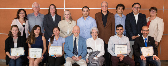
On September 14, 2015, the UCLA-DOE Institute of Genomics and Proteomics will host a 1-day minisymposium on Frontier Problems and Technologies in Bioenergy and Biodesign. The symposium aims to expose the UCLA campus and nearby research communities to important new energy-related research and technology developments. A number of leading investigators will discuss their latest work.

Schedule

(All activities except lunch will be in Boyer Hall 159)

8:30 – 9:00 Meet and greet — coffee and pastries

9:00 – 9:30 Introductory remarks (UCLA – Kelsey Martin 9:00-9:10;
DOE – Roland Hirsch 9:15 – 9:25)

9:30 – 10:00 Intro to UCLA-DOE Institute activities (Sabeeha and Todd)

SESSION ON EMERGING TECHNOLOGIES:

10:00 – 10:25 James Evans (PNNL, EMSL)

10:30 – 10:55 Petra Fromme (ASU)

11:00 – 11:25 Xijie Wang (SLAC)

11:30 – 12:00 – Posters and discussions

12:00 – 1:00 – Lunch

SESSION ON MICROBES AND PLANTS:

1:00 – 1:25 Sabeeha Merchant

1:30 – 1:55 Ken Kemner (DOE, Argonne)

2:00 – 2:55 Chentao Lin (UCLA)

2:30 – 3:00 coffee break

SESSION ON BIODESIGN:

3:00 – 3:25 Ron Zuckermann (LBNL)

3:30 – 3:55 Danielle Tullman-Ercek (Berkeley)

4:30 – 5:15 Roundtable discussions – DOE visitors with DOE PI’s

Break

5:45 Depart for dinner

6:00 Dinner

Todd Yeates has received The DeLano Award for Computational Biosciences

Todd Yeates has received The DeLano Award for Computational Biosciences by the American Society for Biochemistry and Molecular Biology (ASBMB). The Award is given in the field of computational biology for “the most accessible and innovative development or application of computer technology to enhance research in the life sciences at the molecular level.”

The prize was established in memory of Warren Lyford DeLano, who developed PyMOL, an open source molecular viewer software and was an advocate for open source practices in the sciences.

Previous winners of this prestigious award include Vijay Pande, Michael Levitt, Helen Berman, Barry Honig and Axel Brunger.

Todd is recognized for his multiple, profound contributions to computational biology. These include: methods to design large, open protein shells capable of encapsulating cargo; methods to infer protein interactions from genome sequences; a powerful method to detect errors in protein structures; and a method to detect twinning which often bedevils protein structure determination.

See this article at the UCLA Newsroom

http://services.mbi.ucla.edu/DiffAcc

An illustration of ‘diffusion accessibility’ (Tsai, Holton, and Yeates, Protein Sci. 2015 Jul 16) a computational method for characterizing the surface shapes and binding clefts of macromolecules, implemented and rendered in PyMol. [http://services.mbi.ucla.edu/DiffAcc]

UCLA-DOE Institute to support UCLA’s iGEM team (International Genetically Engineered Machines) in the 2015 annual competition in Boston.


The main program at the iGEM Foundation is the International Genetically Engineered Machine (iGEM) Competition. Click here for more information.

See the video made by the UCLA iGEM undergraduate team

Igemlogo_officiallogo

Next APS Trip

October 21, 2015